The development of chick embryos has been studied since Aristotle. It is one of the most intensely studied organisms. One reason for this is that there are great similarities between avian and mammalian embryology. Another advantage is that the embryo is relatively accessible and can be operated on or treated with teratogens (substances that perturb development) while still in ovo(in the egg). Finally, fertilized chicken eggs are available year round and can be "held" at 10-14oC for several weeks before being set, producing embryos upon demand.
Embedded embryos
1. Examine plastic embedded specimens of "13-hour" or "16-hour", "24-hour", "33-hour","48-hour" and "72-hour" chick embryos. These terms refer to classic studies of chick development. In reality, chick embryos will develop at different rates for a number of reasons, including incubation temperature. In modern times, chicks always take longer to reach these stages. Two more accurate ways of describing the relative developmental age of a chick embryo are (1) the number of somites and (2) comparison to a staging series, such as the one devised by Hamburger and Hamilton (H&H; hamburger.pdf). The somites are the blocks of tissue on either side of the neural tube. Try to determine the number of somites and the H&H stage number of your embryos.
2. Try to identify the structures indicated in the diagrams. Pay particular attention to the developing circulatory system. Try to determine which germ layer forms each structure. Highlights include:
13- to 16-hour.Identify the area opaca and area pellucida. Locate the primitive streak. Hensen's node is located at the anterior end of the streak.
24-hour.Locate the neural folds and Hensen's node. Identify the anterior end of the embryo.
33-hour.The embryo is lying along the center of the blastodisc, dorsal side up. The heart is to the right, along the side of the hindbrain. It is a simple, looped tube;connected to the vitelline vein and the ventral aorta. What is the function of the vitelline vein? Identify the forebrain, midbrain and hindbrain. The optic vesicles are outpockets on either side of the forebrain. The neural plate runs along the dorsal midline and is not yet closed. You may also be able to see the notochord running along the midline.
48-hour.The head has bent forward and the body has begun to twist. The optic cup(eye) and the otic vesicle (ear) have formed. The neural tube has closed, except in the region of the hindbrain. A series of aortic arches run between the pouches
in the pharynx and converge into the dorsal aorta. The heart has begun to differentiate into an atrium and a ventricle.
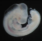
72-hour.The flexure of the embryo is now pronounced and the embryo is lying on
its left side. The brain has further subdivided and the olfactory pit (nose) is
present over the tip of the forebrain. The lens of the eye should be visible.
Lateral swellings representing the limb buds and a curved tail are also present.
Living embryos
1. Isolate a living 3- to 6-day chick embryo. Obtain an egg and clean scissors, forceps,
a plastic spoon. First prepare a small petri dish withHoward Ringer's solution.
Gently rotate egg, clean with 70% ethanol and set down for a few minutes to
allow the embryo to float to the top. Open up the blunt end of the shell and
carefully peel back the shell membranes. Observe the embryo under the
dissecting scope.
2. Measure the heart rate and observe any other movement. Grab
the outer ring of the blastodisc near embryo with fine forceps in your non-cutting
hand. Try not to pinch any blood vessels. Quickly cut around embryo.
3. Keeping hold of the embryo with the forceps, pick up a plastic spoon with your
cutting hand and slide it under the embryo. Continue to hold onto embyo, lift
with the spoon and transfer to the petri dish filled with Howard's and examine
with the dissecting microscope. have your lab partner stand by with scissors in
case the embryo wasn't completely detached. If the dish is cloudy with yolk,
transfer embryo to a fresh dish of Howard's.
Note: an alternate method of embryo isolation is to break the egg open into a finger bowl of Howard's. This is similar to cracking an egg for culinary purposes, but the yolks of warm eggs are more fragile than those of refrigerated ones. First, place the egg in a horizontal position to allow the embryo to float to one side. Next, keeping the same orientation, crack the egg gently against the side of a finger bowl filled with Howard's. Submerge the egg and gently pull the ends appart to crack the egg open.

4. Isolate a 1- or 2-day embryo. Clean your dissecting equipment and prepare a fresh
dish of Howard's. Clean an egg and allow the embryo to float to the top.
Open the blunt end of the egg and remove the shell membranes. the embryo
may not be visible to the naked eye. The blastodisc is located above a small ring
of white yolk. Remove albumin with wide-mouth pipet and shell with forceps.
5. Drop a filter paper disc around your embryo. Hold on to the filter 6. Examine both embryos. Pay particular attention to the heart and circulation, and 7. For each embryo,determine the H&H stage. How do these embryos compare to 8. Clean your instruments well with warm water, distilled water and 70% ethanol.
paper with fine forceps and cut around the ring with your sharp scissors.
Transfer the embryo to a small petri dish with Howard's Ringer's solution.
to the developing neural tube. Compare the heart rate between the 2 embryos.
Which side was towards the yolk? The reddish spots on the 2-day blastodisc are
the blood islands, the sites of hematopoesis. The embryo is covered with a clear
protein layer known as the vitelline membrane. This may start to peel away
from the embryo.
the stained specimens?
Dry before returning to case. Discard the shells and the yolk/albumin remains.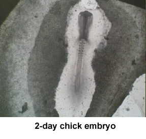
3-day chick embryo
蛋白質作為構成人體組織器官的支架和主要物質,在人體生命活動中起著重要作用。蛋白質的相互作用能產生許多效應,如形成特異底物作用通道、生成新的結合位點、失活、作用底物專一性和動力學變化等,細胞的代謝、信號......
2021年9月9日,無錫臻和生物科技有限公司(以下簡稱“臻和科技”)與美國VyantBio公司簽署TissueofOrigin?(以下簡稱“TOO?”)全球權益和ZL轉讓協議,全資收購這款唯一獲FDA......
2021年7月20日,JournalofCellularPhysiology及JournalofCellularBiochemistry同時撤回了中國學者49篇文章。從2019年開始,Journalo......
磷酸甘油酸突變酶1(PGAM1)通過其代謝活性以及與其他蛋白質(例如α平滑肌肌動蛋白(ACTA2))的相互作用,在癌癥代謝和腫瘤進展中起關鍵作用。變構調節被認為是發現針對PGAM1的高選擇性和有效抑制......
磷酸甘油酸突變酶1(PGAM1)通過其代謝活性以及與其他蛋白質(例如α平滑肌肌動蛋白(ACTA2))的相互作用,在癌癥代謝和腫瘤進展中起關鍵作用。變構調節被認為是發現針對PGAM1的高選擇性和有效抑制......
2018年12月6日,來自圣迭戈的消息——Illumina公司(納斯達克股票代碼:ILMN)今天宣布推出新型高密度基因分型芯片Infinium?GlobalDiversityArray。這款芯片設計源......
SDS-PAGE異常電泳現象及分析SDS-PAGEHallofShame.pdf 很不錯的東東~~推薦下~......
Preparationofdenaturing6%polyacrylamidegelsformicrosatelliteanalysis(alsoforSSAP,high-resolutionIRAP......